
24 X 7 online support
skcfindivar@gmail.com
Screenings and examinations performed during pregnancy may provide valuable information about the state of the unborn child's health before the infant is brought into the world. Screening tests are often performed in both the first and second pregnancy trimesters to evaluate whether or not there are any potential health problems to the unborn child.
Most of the early-stage examinations are noninvasive methods.
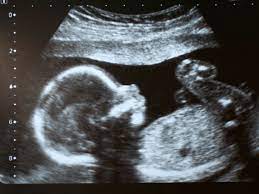
Nuchal translucency, often known as NT, is a deposit of fluid that develops underneath the skin at the nape of a fetus's neck.
To some degree, fluid will always be present in the back of a developing baby's neck. However, many newborns with Down syndrome have an elevated quantity. Because of this, the NT scan is a diagnostic tool used to screen for Down syndrome.
Additionally, during the first trimester, doctors check for the fetal nasal bone (NB). The chance of chromosomal problems rises if no nasal bone is visible in the growing fetus.
Trisomy 21 and other large aneuploidies may be found in roughly 80% of pregnancies by NT screening, with a 5% false positive rate. A 90 percent improvement in detection is made by combining NT, maternal serum-free hCG, and PAPP-A. There is no proof that by looking at the nasal bone, ductus venous flow, and tricuspid flow, the detection rate may rise to roughly 95%, reducing the false positive rate to 3%.
An ultrasound called an NT scan is performed during the first trimester to assess the fetus's chances for chromosomal abnormalities, including down syndrome. Typically, a blood test is also conducted. You may decide to get a CVS or amnio for a clinical diagnosis if the screen shows that the fetus may have an issue. In addition to screening for trisomy 21 (Down syndrome), the NT scanning will check for:
The scan is also often referred to as the Fetal Anomaly Scan. As the name implies, this scan is conducted to see whether the fetus is developing typically. Identifying any congenital disabilities in the developing baby makes it one of the most crucial scans performed during pregnancy. This scan is performed between weeks 18 and 22 of pregnancy. The infant is examined from head to toe to check for any anomalies during this scan.
A TIFFA scan may provide several important information, including:
Screening for genetic conditions during pregnancy may detect any possible genetic risks or problems before the baby is born. Currently, a screening test for Mongolism does not include invasive procedures and has been shown to be more accurate than previous methods.
A condition known as Down syndrome results when a fetus has an additional copy of chromosome 21. This condition is sometimes referred to as trisomy 21. Children born with an extra copy of chromosome 21 suffer mental impairment, the physical manifestations of Down syndrome, and around a forty percent chance of developing major cardiac issues. As early as 10 weeks of pregnancy, screening tests may establish whether or not there is a possibility of having a kid with a genetic disease such as Down syndrome.
If the screening test is positive, further invasive diagnostic tests are carried out, including a CVS test during 10 to 13 weeks of pregnancy and amniocentesis during the 15th week of pregnancy.